


What is Brain Tumour Surgery?
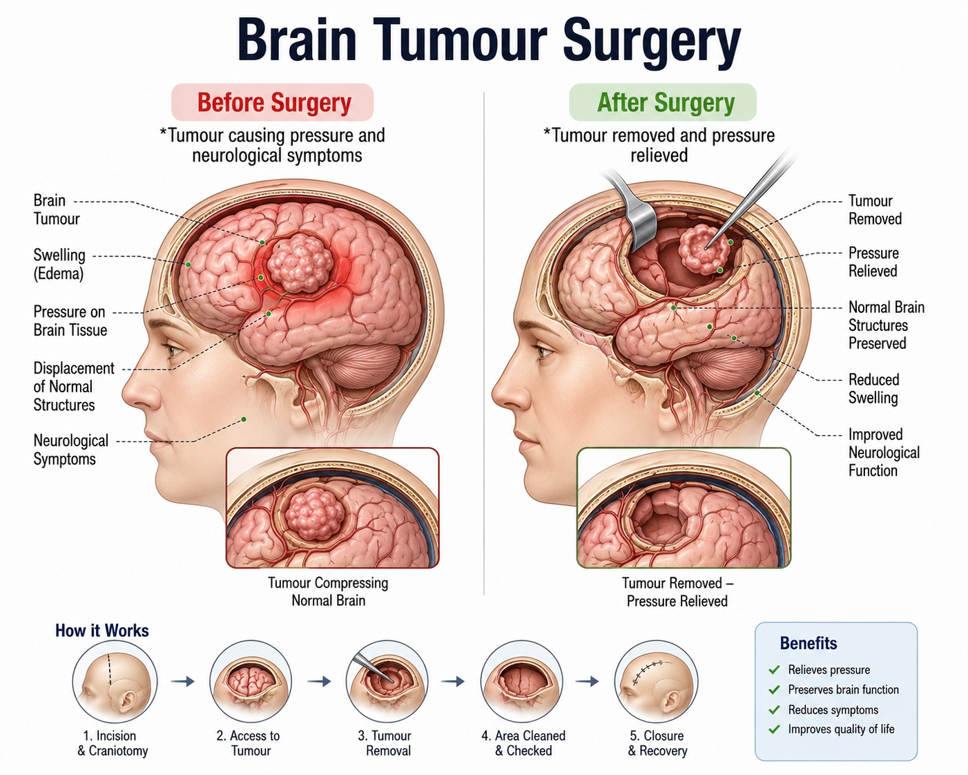
Brain tumour surgery, primarily performed via craniotomy, involves opening the skull to safely locate and resect (remove) abnormal tissue growths within or around the brain. The primary goals are maximal tumour resection, relief of intracranial pressure, tissue biopsy for accurate histopathological diagnosis, and protection of speech, motor, and cognitive pathways.
Who Needs Brain Tumour Surgery?
Surgery is indicated for both primary brain tumours (originating in brain tissue or meninges) and metastatic lesions (spreading from elsewhere in the body). Treatment decisions are made based on tumour location, size, growth rate, and patient symptoms.
Key clinical indicators include:
- Persistent, worsening headaches accompanied by morning nausea or vomiting.
- New-onset seizures or focal motor weakness (e.g., facial drooping or weakness on one side).
- Cognitive changes, memory loss, visual field defects, or personality shifts.
- MRI or CT evidence of space-occupying intracranial mass causing mass effect or brain shift.
- Hydrocephalus caused by tumour obstruction of fluid pathways.
Types of Brain Tumour Surgery
- Craniotomy: Removal of a section of bone (bone flap) to access and resect the tumour, after which the bone is secured back in place.
- Awake Craniotomy: Performed while the patient is conscious during mapping steps to monitor motor or speech function in real time when tumours lie near eloquent brain areas.
- Endoscopic Endonasal Approach: Minimally invasive removal of pituitary and skull-base tumours through the nasal passages without skin incisions.
- Stereotactic Biopsy: Minimally invasive needle sampling of deep-seated or inoperable tumours using 3D image guidance.
Note on Technology: Neuronavigation (GPS for the brain) and intraoperative MRI/ultrasound allow surgeons to map functional boundaries and maximize safe tumour removal.
Benefits of Brain Tumour Surgery
- Immediate reduction of intracranial pressure and mass effect.
- Definitive tissue acquisition for molecular profiling and targeted therapies.
- Potential for complete cure in benign tumours (e.g., meningiomas).
- Improved survival rates and symptom control in malignant brain tumours.
What to Expect Before Surgery?
Preparation ensures surgical safety and detailed anatomical mapping:
- Advanced Neuroimaging: High-resolution contrast MRI, functional MRI (fMRI), and diffusion tensor imaging (DTI) tractography.
- Medical & Neurological Evaluation: Assessment of baseline motor, sensory, and speech functions, alongside routine cardiac and blood panels.
- Steroid Administration: Pre-operative corticosteroids (e.g., dexamethasone) to reduce peritumoral brain swelling.
- Anticonvulsant Management: Prophylactic anti-seizure medication if epilepsy risk is high.
Complete Procedure
The surgical process typically follows these steps:
- Administration of general anesthesia (or scalp block with conscious sedation for awake procedures).
- Rigid head fixation in a Mayfield frame to prevent movement and integrate with neuronavigation systems.
- Incision of the scalp and creation of a bone flap (craniotomy).
- Opening the dura mater (outer membrane covering the brain).
- Microscopic or endoscopic dissection to resect the tumour tissue while sparing healthy brain tissue and blood vessels.
- Hemostasis verification and dural closure (often using dural grafts or sealants).
- Replacing the bone flap with titanium plates and screws, followed by scalp closure.
What to Expect During Surgery?
The procedure usually lasts between 3 and 7 hours depending on complexity. Operating room teams include neurosurgeons, neuro-anesthesiologists, surgical techs, and neurophysiologists monitoring nerve activity continuously.
Navigation-Guided vs Traditional
| Feature | Traditional Craniotomy | Neuronavigation & Intraoperative Mapping |
|---|---|---|
| Surgical Precision | Relies primarily on anatomical landmarks. | Sub-millimeter real-time tracking with fused MRI datasets. |
| Eloquent Area Safety | Higher risk near speech/motor zones. | Intraoperative neuro-monitoring and awake mapping minimize risk. |
| Resection Completeness | Visual estimation of margins. | High margin precision, often combined with fluorescence guidance (5-ALA). |
| Recovery Profile | Standard neurological recovery. | Optimized recovery with reduced operative trauma. |
Success Rate and Safety
Surgical outcomes depend on tumour grade and location. Benign tumour resection carries a high long-term success rate (>90% long-term control). For high-grade gliomas, maximal safe resection significantly enhances the effectiveness of post-operative radiation and chemotherapy.
Brain Tumour Surgery Cost Estimate
| Type of Procedure | Conventional Surgery (INR) | Navigation/Advanced Guided (INR) |
|---|---|---|
| Standard Craniotomy & Resection | ₹3,50,000 - ₹5,50,000 | ₹4,50,000 - ₹7,00,000 |
| Awake Craniotomy / Intraoperative Monitoring | ₹4,50,000 - ₹6,50,000 | ₹5,50,000 - ₹8,50,000+ |
| Endoscopic Skull Base / Pituitary Surgery | ₹3,50,000 - ₹6,00,000 | ₹5,00,000 - ₹9,50,000 |
Disclaimer: Estimated baseline costs. Final charges vary based on ICU duration, histopathology requirements, intraoperative imaging, and hospital category.
Recovery Guide & Post-Operative Care
Recovery Timeline
- Day 0-2: Intensive Care Unit (ICU) monitoring; frequent neurological checks.
- Day 3-7: Transition to neuro ward; physical and occupational therapy initiation.
- Week 2-4: Scalp suture/staple removal; pathology review to decide adjuvant therapies (radiation/chemotherapy).
- Month 2-6: Gradual cognitive and physical rehabilitation; return to routine daily activities under clearance.
Post-Op Precautions & Care
- Seizure Precautions: Strict compliance with prescribed anti-epileptic medications.
- Wound Care: Keep scalp incisions clean and dry; watch for swelling or clear fluid discharge (CSF leak).
- Activity Restrictions: Avoid heavy lifting, bending, or strenuous exertion for 6-8 weeks.
- Follow-up Imaging: Scheduled baseline MRI within 24-72 hours post-surgery, followed by periodic surveillance scans.
Frequently Asked Questions
How long does recovery take after brain tumour surgery?⌄
Initial hospital recovery takes 5 to 10 days. Full recovery and rehabilitation take anywhere from 6 weeks to several months depending on the tumour size, location, and whether secondary treatments like chemotherapy are needed.
Will my entire head be shaved for brain surgery?⌄
No, modern neurosurgical techniques only shave a small strip of hair directly along the planned incision line, preserving as much hair as possible.
Is awake brain surgery painful?⌄
No, the brain tissue itself has no pain receptors. Local anesthetics block feeling in the scalp, and sedation keeps the patient relaxed and comfortable throughout the procedure.
What are the main risks of brain tumour surgery?⌄
Risks include infection, bleeding, brain swelling, seizures, and localized neurological deficits (such as temporary or permanent weakness or speech difficulty), which are minimized through advanced surgical navigation.
When can I return to work or driving?⌄
Most patients can return to light work in 6 to 12 weeks. Driving restrictions depend on seizure history and local legal regulations, typically requiring clearance from your neurologist.