










What is Retinal Detachment Surgery?
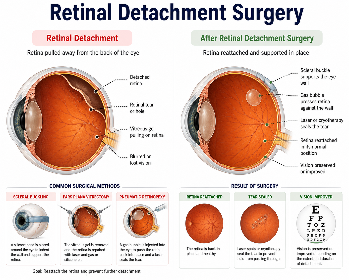
Retinal Detachment Surgery encompasses urgent specialized surgical techniques—primarily Pars Plana Vitrectomy (PPV) and Scleral Buckling—designed to reattach the neurosensory retina to the underlying retinal pigment epithelium (RPE). When fluid passes through a retinal tear, it separates the retina from its blood and nutrient supply. Without prompt surgical reattachment, oxygen deprivation leads to permanent loss of photoreceptor cells and irreversible blindness.
Who Needs Retinal Detachment Surgery?
Immediate surgical intervention is required whenever a retinal tear, traction, or detachment is diagnosed by an ophthalmologist.
Key clinical indicators include:
- Sudden appearance of numerous floaters (specks or cobwebs) in vision.
- Flashes of light (photopsia) in one or both eyes, especially in peripheral vision.
- A dark shadow or "curtain" falling across the field of vision.
- Sudden blurring or loss of central visual acuity.
- History of severe myopia, ocular trauma, or prior cataract surgery complications.
Surgical Techniques for Retinal Detachment
- Pars Plana Vitrectomy (PPV): Micro-incisional removal of the clear vitreous gel pulling on the retina, flattening the retina with air/gas/oil, and sealing tears with endolaser.
- Scleral Buckling: Securing a tiny flexible silicone band or sponge around the outer wall of the eye (sclera) to indent it inward against the detached retina.
- Pneumatic Retinopexy: In-office injection of an expandable gas bubble into the vitreous cavity accompanied by cryopexy/laser, suitable for uncomplicated upper retinal tears.
- Combined Vitrectomy & Buckling: Used for complex, recurrent, or tractional retinal detachments.
Note on Tamponade Agents: Intraocular Gas (SF6 or C3F8) or Silicone Oil is injected into the eye to hold the reattached retina in place during healing.
Benefits of Retinal Detachment Surgery
- Immediate prevention of total, permanent blindness in the affected eye.
- Reattachment and structural stabilization of the neurosensory retina.
- Potential for substantial visual recovery, especially if performed before macula off.
- Long-term preservation of peripheral visual field and eye integrity.
What to Expect Before Surgery?
Preoperative evaluation is conducted urgently upon presentation:
- Dilated Fundus Examination: Indirect ophthalmoscopy to identify all retinal tears and detachment boundaries.
- B-Scan Ocular Ultrasound: Sound wave imaging used when dense vitreous hemorrhage obscures optical view.
- OCT Macula Scan: High-resolution cross-sectional scan to determine if the central macula is attached or detached.
- General Anesthesia Clearance: Fasting and medical checks for urgent surgery.
Complete Procedure
The step-by-step process for Pars Plana Vitrectomy involves:
- Administration of local block or general anesthesia to ensure complete pain relief and eye immobility.
- Making 3 micro-gauge cannula trocars (23G, 25G, or 27G) through the pars plana region.
- Inserting a fiber-optic light source, infusion line, and micro-vitrector tool.
- Removing the vitreous gel and releasing all traction pulling on the retina.
- Re-expanding and flattening the retina using heavy liquid (Perfluorocarbon) or air.
- Applying Endolaser photocoagulation or Cryopexy around all retinal tears to weld them shut.
- Exchanging fluid for a intraocular gas bubble or silicone oil tamponade.
What to Expect During Surgery?
Surgery duration ranges from 1 to 2.5 hours depending on complexity. You will be fully monitored under local block with sedation or general anesthesia.
Vitrectomy vs Scleral Buckle vs Pneumatic Retinopexy
| Feature | Pars Plana Vitrectomy | Scleral Buckle | Pneumatic Retinopexy |
|---|---|---|---|
| Procedure Site | Inside the eye cavity | Outside the eye wall | Office gas injection |
| Tamponade Used | Gas bubble or Silicone Oil | External silicone band | Expandable gas bubble |
| Posturing Required | Face-down posturing needed | Minimal posturing | Specific head positioning |
| Best For | Complex/posterior tears | Young patients/phakic eyes | Uncomplicated superior tears |
Success Rate and Safety
Anatomical reattachment of the retina is achieved in 85% to 90% of cases in a single surgery, and over 95% with secondary intervention. Visual recovery depends heavily on whether the central macula was involved prior to surgery.
Retinal Detachment Surgery Cost Estimate
| Procedure Type | Cost Range per Eye (INR) |
|---|---|
| Standard Scleral Buckling Surgery | ₹45,000 - ₹75,000 |
| Micro-Incisional Vitrectomy (23G/25G) | ₹65,000 - ₹1,10,000 |
| Complex Vitrectomy + Silicone Oil Insertion | ₹85,000 - ₹1,40,000 |
| Secondary Silicone Oil Removal Surgery | ₹25,000 - ₹45,000 |
Disclaimer: Final cost varies based on surgical duration, tamponade material used, ICU/stay requirements, and surgeon expertise.
Recovery Guide & Post-Operative Care
Recovery Timeline
- Week 1-2: Strict prone (face-down) positioning if gas/oil was used; vision is obscured by gas bubble.
- Week 3-6: Gas bubble slowly absorbs naturally (if gas used); visual clarity progressively improves.
- Month 3-6: Complete tissue healing; silicone oil removal planned if oil was instilled.
Post-Op Precautions & Care
- Prone Head Posturing: Maintaining face-down positioning for 5–10 days as instructed by the surgeon is critical for success.
- NO AIR TRAVEL: Absolute prohibition of air travel or high-altitude trips while gas bubble is inside the eye (risk of extreme pressure elevation and blindness).
- Eye Protection: Wear a rigid eye shield, especially during sleep.
- Hygiene & Drops: Antibiotic and anti-inflammatory eye drops applied diligently.
Frequently Asked Questions
Why is face-down posturing necessary after retinal surgery?⌄
Face-down positioning allows the intraocular gas bubble or silicone oil to float upward and press directly against the repaired retinal tear, sealing it in place while it heals.
Can I fly on an airplane after retinal detachment surgery?⌄
NO. If a gas bubble was used, flying is strictly prohibited because atmospheric pressure changes cause the gas bubble to expand rapidly, causing severe eye pain and irreversible blindness. Flying is safe only after the gas completely absorbs or if silicone oil was used.
Will my vision return to 100% after retinal surgery?⌄
Visual recovery depends on whether the central retina (macula) was detached prior to surgery. If the macula remained attached, visual recovery is excellent; if detached, vision improves significantly but may not reach 100% baseline.
How long does the gas bubble remain inside the eye?⌄
Depending on the gas type used (SF6 or C3F8), the bubble naturally absorbs and is replaced by natural eye fluid over 2 to 8 weeks.
When is silicone oil removed after vitrectomy?⌄
Silicone oil does not dissolve naturally and is safely removed via a minor secondary surgical procedure typically 3 to 6 months after the initial retina reattachment.